

Pregnancy and giving baby birth to a fetus are the beautiful and sweet moments on the face of the earth but may become sometimes painful and frightening processes involving different hardships. However, it is necessary to monitor the happenings while giving birth to a fetus in a safest and controlled way.
How to read a contraction monitor to stay updated has been frequently asked. Different devices are used and the process is termed is Cardiotocography, shortly CTG to read and time the contraction and baby heart rate. In order to avoid any unforeseen circumstances people often search how to read a contraction monitor. Here is a guide for you to learn all the ways of reading a contraction monitor.
Table of Contents
- What is Cardiotocography (CTG)?
- Determining If the Pregnancy Is High or Low Risk
- Continuous Monitoring and High-Risk Pregnancy
- Contraction Monitors – Types & Uses
- Key Terms for Uterine Contractions
- Interpreting an Electronic Contraction Monitor
- The Graphs on Monitor Screen
- Normal Contractions
- Types of Variability
- Accelerations and Decelerations
- Interventions
- Conclusion
- Frequently Asked Questions
What is Cardiotocography (CTG)?
Cardiocartography, shortly as CTG, is used for fetal monitoring and is helpful in detecting the fetal heart rate and uterine contractions while delivery or at the time of baby birth. It is used to examine the fetal as well as the mother, nurses or doctors try to know the situation of the fetal that wither his/her heart rate is normal or not. Thus, it becomes crucial to know reading a contraction monitor.
For mother, it is used to monitor the uterine contraction that wither the contractions are normal or not, if not normal then they inject some hormones etc. It is often used for the early examination of the fetal. If the CTG is normal the doctors advise the time of the delivery while in the unpleasant situations there is a dire of careful observations.
Similar Reading: How to Self Soothe Anxious Attachment
Reading and Interpreting a CTG
Reading a CTG is not everyone’s cup of tea, it needs to understand the basic terminologies and the characters used for them. The common acronym used everywhere is DR C BRAVADO, with the help of which you will be able to not only read but also interpret the CTG.

Determining If the Pregnancy Is High or Low Risk
The doctors and nurses use equipment according to the patients’ situations, if there is no risk and abnormality they go with the one and for the very close observations in the risky and complicated delivery they opt another.
There may take place issues of water break, the fetal position may displace or there are chances of other numerous abnormalities. So, the most important thing to be taken into consideration is the determination of patient situation, for either cases there are different monitors and different methods of monitoring.
Continuous Monitoring and High-Risk Pregnancy
The continuous monitoring is used for the high risk pregnancy because it needs to be examined for every second and minutes. There are some major reasons behind this fact, if mother is feeling any fetal distress in the labor, if mother is anesthetic to induce the labor pain. It can be also used mother has any issue like hypertension or if she is diabetic.
If a patient had delivery with cesarean section she is most likely to face complication and should be monitored with continues monitoring. It can also be used if the doctor wants to take extra care of the patient, so using this does not necessarily mean that there is complications.
Contraction Monitors – Types & Uses
Typically, two types of contraction monitors have been used by the medical practitioner to find out and determine the stage of pregnancy and also for the diagnosis of any pre mature issues. These are also used in fetal monitoring, continuous electronic fetal monitoring or structured intermittent auscultation Know types are as under: -
a). Continuous, Or Electronic
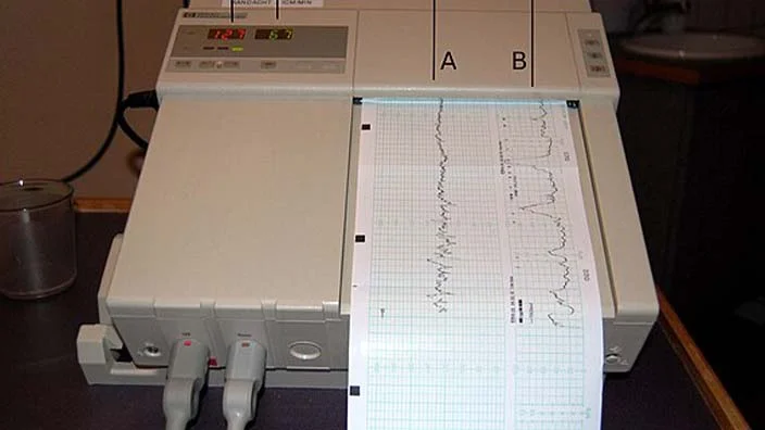
A continuous contraction monitor is also termed an electronic contraction monitor. It is widely used in normal situations generally, detects and shows the fetal heart rate as well as the mother’s uterine contractions on a screen of monitor.

The continuous or electronic fetal monitor is often used externally, an ultrasound transducer, which is also known as tycodynamometer or TOCO in short, is placed on mother’s belly to detect the fetal heart rate. The second sensing monitor is strapped on the top of mother’s abdomen to examine the uterine contractions.
To use the electronic monitor internally, which is rarely used, an Intrauterine Pressure Catheter, in short IUPC, is used for the strength of mother’s contractions. An electrode of small size is placed inside the vagina of Mother and carefully fixed to the scalp of fetus for monitoring the heart rate. The mother’s uterine contractions will also be shown on the screen if she is not aware of them on account of anesthesia. The contractions will appear on the screen as a pointed peak like structure.
b). Intermittent or Auscultation
The second type of fetal and contraction monitoring is Intermittent or Auscultation, measures the periodic contractions rather than constant. It is a kind of manual monitoring in which the nurse having a particular stethoscope also known as Fetoscope or Doppler Transducer will detect the fetal heart rate by placing the device near mother’s stomach.
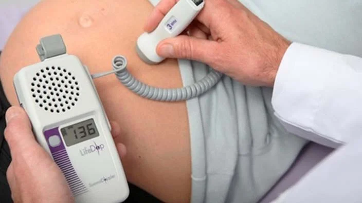
The method is mostly used on account of the ease of moving in the labor. A women can only allowed to move in the low risk pregnancy, so this type of monitoring is preferred in the pregnancy lacking any complications. Without positioning any sensing any electrode mother can change her positions as well. A nurse after every particular interval of time examines the fetal heart rate with her fetoscope.
Key Terms for Uterine Contractions
Here are some key terms for the uterine contractions which bear significant meanings and need to be understood. The first one is frequency which means that how far apart the contractions are. The second one is duration which indicates that how long the contraction last. Third, intensity symbolizes that strength of contractions and the last one, rest bears the meaning of tone and time that the uterus should be fragile enough to palpation for 60 seconds, the minimum limit.
Interpreting an Electronic Contraction Monitor
Interpreting an electronic contraction monitor is very necessary for everyone, not only during the delivery but also it may help you reading your contractions and fetal heart rate at home on the paper. During the fetal monitoring there may take place some complications either with the contraction of mother or with the baby heart rate and in order to avoid such unforeseen circumstances people often search how to read a contraction monitor. Here are the basics of interpreting an electronic contraction monitor.
The Graphs on Monitor Screen
The nurses place two sensing electrodes, an ultrasound transducer, which is also known as tycodynamometer or TOCO in short, is placed on mother’s belly to detect the fetal heart rate. The second sensing monitor is strapped on the top of mother’s abdomen to examine the uterine contractions.
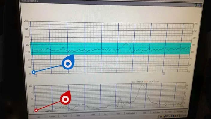
These two are connected with wires to the monitor, the monitor often has two graphs one above the other. The top one exhibits the fetal heart rate in beats per minutes or in short BPM while the one below it shows the mother uterine contractions in mmHg.
a). Monitoring Baby's BPM and Mother’s Uterine Contractions
The monitor is kept besides the hospital beds, there you will see two graphs one over another. The top one exhibits the fetal heart rate in beats per minutes or in short BPM while the one below it shows the mother uterine contractions.

The lines on the graph direct from right to left which indicate that the line starts from the right sides and are the most recent results. The fetal heart rate is lined up with the mother’s uterine contractions. You can also get the printouts of the graphs which you can read in the home later. Anyhow, if the doctor is using electronic monitor he/she may assist you and interpret the graphs.
b). The X-Axis Times Your Contractions And Your Baby's Heart Rate
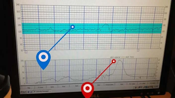
The X-axis is also called the horizontal line is same for both the graphs, shows the time in minutes on both the graphs, one minute is shown between the red and blue indicator. There are 6 sections of 10 seconds between each minute. The lines on both the graphs move along showing uterine contractions and fetal heart rate.
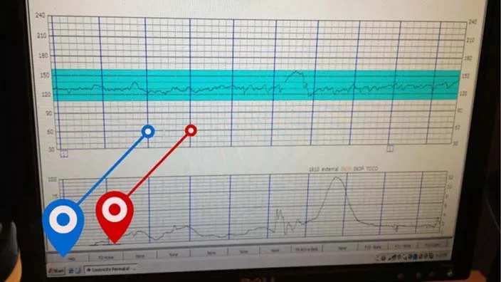
You can easily notice each contraction or heart rate and how both are far apart from the next in one minute. The contractions function as tightening the top of the uterus and apply pressure on the cervix. This pressure dilates the cervix and provides a way to the fetus to move downward. The time interval for each contraction ranges from 30 seconds to 70 seconds and arises usually after 5 to 10 minutes.
c). The Y-Axis of the Fetus’ Chart Displays BPM
The Y-axis or vertical line of the both the graphs are different from each other. The vertical axis on the graph of fetus indicates the fetal heart rate in beats per minutes, shortly BPM. The normal fetal BPM ranges from 110 to 160, there may take place some acceleration or deceleration but nothing to worry, that’s normal.
It may seem frightening to you that the fetus shows abnormal heart beats and the graph may threaten you but it happens in the labor. The BPM is measured in 10 incremental scales which mark every 30 beats.
d). The Y-Axis Mother’s Chart Shows Contraction Strength
The Y-axis or the vertical axis signals the intensity contractions in millimeters of mercury, in short mmHg. The higher number shows that the contractions are strong, so there are a direct relation between them, the high the mmHg the stronger the contractions.
At the very begging of the labor the graph may show the lines ranges from 50 to 70mmHg. It happens when the cervix is expected to dilate from 6 to 10 cm with which the intensity, frequency and regularity of the contractions are supposed to be. The minimum limit of contractions may be recorded at the very moment of giving birth to the fetus when the strength of contractions increases from 80 to 100 mmHg.
Normal Contractions
The contractions will be considered normal with the frequency if these are 2 to 3 minutes apart in active labor. The duration must be 60 seconds which means that the contraction must last for 60 seconds. The intensity of normal contractions will be 60 mmHg and the rest must be for 60 seconds between contractions.
Types of Variability
Throughout the whole process there may occur some variability and can be shown in the graphs by the line. What is that variability and what does it mean? Let’s discuss all of them. The lack of variability means that there is no jiggling and can be considered threatening.
The minimal variability is when the lines become flatter, it does not necessarily mean that there is problem. It may signals that the baby is sleeping or sad and is not yet ready to move. The most desired variability is the moderate which indicates the normality. The marked variability shows the jiggling and can be understood that the baby is stressed.
Accelerations and Decelerations

While giving birth to fetus is a sweet and joyous moment on the face of the earth but sometimes it may be painful and frightening. The graphs may sometimes shows accelerations and deceleration in the lines indicating the surges and declines in the contractions and in fetus heart rate as well. The acceleration and deceleration both become normal within 2 minutes.
The temporary incline in the FHR means the fetus is in dire need of oxygen. However, the decelerations or decels may be fearful sometimes. There are three types of decelerations; early, variable and late decels. The graph with early deceleration shows shallow bowl shaped dips that signals head compression. In the variable deceleration the graph appears sharp V dips and signals out the cord compression. The late deceleration indicated hypoxia, the decreased oxygen.
Interventions
Sometimes the situation may become unpleasant which may need a dire need of interventions. The interventions made to be are simply shown by the acronym as ROADI. The nurse may need to reposition the mother. If there is need of oxygen, it may be passes via facemask. In severe conditions alert the HC. If there is no need just discontinue oxytocin and give tocolytics to the mother and also increase the IV fluids.
Conclusion
During labor when mother gives birth to the fetus doctors use some devices to monitor the fetal heart rate and mother’s uterine contraction, thus need to know how to read a contraction monitor has gained significance. There are two major types of contraction monitors used in fetal monitoring, Continuous Electronic Fetal Monitoring or structured Intermittent Auscultation.
The nurses place two sensing electrodes, an ultrasound transducer is placed on mother’s belly to detect the fetal heart rate. The second sensing monitor is strapped on the top of mother’s abdomen to examine the uterine contractions. . The normal fetal BPM ranges from 110 to 160 and contractions will be considered normal with the frequency if these are 2 to 3 minutes apart in active labor.
The duration must be 60 seconds which means that the contraction must last for 60 seconds. The intensity of normal contractions will be 60 mmHg and the rest must be for 60 seconds between contractions.
Frequently Asked Questions
How do you read contraction monitor numbers?
The X-axis is also called the horizontal line is same for both the graphs, shows the time in minutes on both the graphs, one minute is shown between the red and blue indicator. The vertical axis on the graph of fetus indicates the fetal heart rate in beats per minutes, shortly BPM and the Y-axis or the vertical axis signals the intensity contractions in millimeters of mercury, in short mmHg.
How do you read a contraction monitor sheet?
A contraction monitor sheet is read while keeping the graphs into consideration. The top one exhibits the fetal heart rate in beats per minutes or in short BPM while the one below it shows the mother uterine contractions. The lines on the graph direct from right to left which indicate that the line starts from the right sides and are the most recent results.
What do braxton hicks look like?
You can easily determine from the signs and symptoms if you are facing Braxton hicks. The upper abdomen of the belly will get harder from the front. You will also observe that the belly will tend to spread a little to the downwards and the will look pointed. Sometimes when the fetus is pressing the belly outward people may also misunderstood the situation.
How far apart should contractions be?
The contractions will be considered normal with the frequency if these are 2 to 3 minutes apart in active labor. The duration must be 60 seconds which means that the contraction must last for 60 seconds. The intensity of normal contractions will be 60 mmHg and the rest must be for 60 seconds between contractions.